

Transmission Electron Microscopy of Control Cultures

All of the content that is put out on this Substack is going to be for free. If you feel so inclined to donate or Sign up for a Paid Subscription that is very much appreciated. It will keep me writing, putting out content and continuing the largest Control Studies Project falsifying Virology.
Here is the report, as we received it after instructing an Independent CRO to conduct Transmission Electron Microscopy of one of the control cultures. The names of the labs involved have been redacted to keep them anonymous (as there are numerous evil people out there who wish to harass and threaten these individuals because they don’t like the Scientific results.)


IMAGES
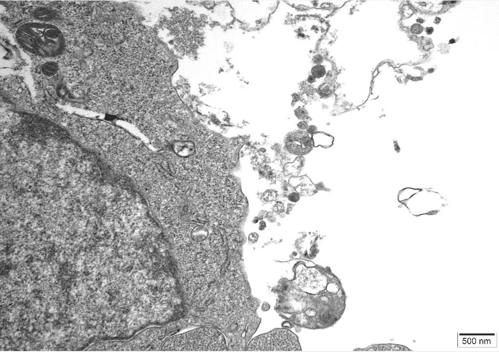
These are the 9 unadulterated images supplied by the CRO.
1.

2.

3.
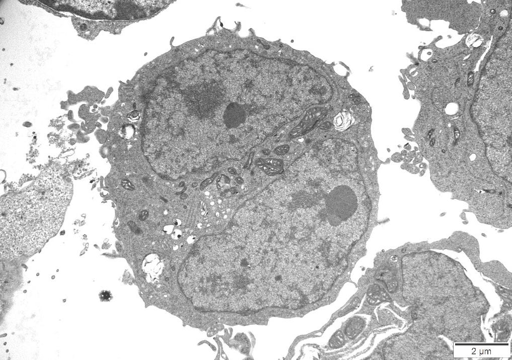
4.

5.
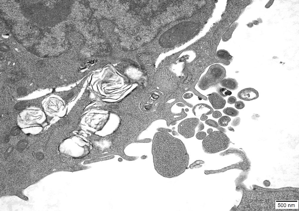
6.

7.

8.

9.
IMAGES OF VIRAL-LIKE PARTICLES AND CROSS-REFERENCED IMAGES
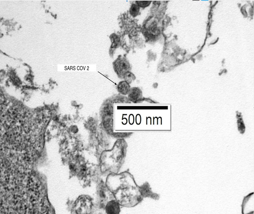
SARS COV 2
From Image 9. A particle of exactly the same size, shape and protein inclusions as the CDC image below of Sars Cov 2, Omicron BA.2.

CDC image of Sars Cov 2.

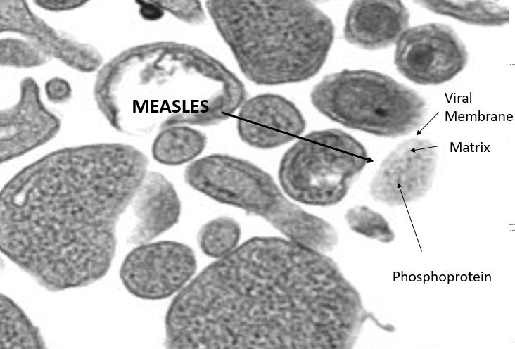
MEASLES
From Image 5. A particle of exactly the same size, shape and protein inclusions as the CDC image below of Measles Virus.


HIV
From Image 9. A particle of exactly the same size, shape and protein inclusions as the CDC image below of HIV Virus.


CONCLUSION
The brief given to the CRO was to look for Extra Cellular Vesicles in the Control Cultures. These were Identified and measured in Images 7&8. They are clearly empty vesicles completely different from the filled particles we have identified and cross-referenced in the rest of the images.
Taking the images, sizes, proteomic description and characteristics of what the CDC considers to be Sars Cov 2, Measles virus and HIV we were able to cross-reference exactly all of these defining features in particles in our control cultures.
Particularly the size and overall shape are two distinctive features which were positively identified and particularly in the case of Measles Virus there is very little explanation as to what else these particles could be, when found in uninfected cultures.
The CRO had total control of what images to take, at no point were they instructed to look for viral-like particles, just images of Extra-Cellular vesicles as we didn’t want to insert any bias into our findings. Despite this, in just 7 images that were not Extra Cellular Vesicles, we manged to positively identify 3 exact matches to distinctive viruses. This was the first Microscopy experiment we carried out as part of the Control Studies, thus the findings that we have are extremely strong. This suggests that if we were to conduct more experiments of this nature but had control over where to image in the cell line we would add to these findings innumerably.
The findings of this paper provide strong evidence that the particles that are being labelled as viruses are clearly either not pathogenic or are misinterpreted cellular debris from the breakdown of the cell-line in starvation medium (Displayed in the Control Culture Isolation Paper).
Further suggestions
There are numerous papers suggesting that tubules within, particularly Renal Cells are the exact size and shape of Sars Cov 2. None more so than in the paper Appearances Can Be Deceiving - Viral-like Inclusions in COVID-19 Negative Renal Biopsies by Electron Microscopy . This sets up some further investigation as to whether these particularly Sars Cov 2 particles maybe found in non-renal uninfected cultures.
There is an obvious call to study more cell-lines with specific regards to Transmission Electron Microscopy for finding potentially many more varieties of viral-like particles. If all or most of the so-called pandemic causing viruses can be located in uninfected cultures they must surely be disregarded as physical entities, or even existing at all.

But Jamie, you did discover viruses, they're just man-made. Maybe dormant, maybe pseudoviruses, maybe foamy viruses like a beer head, but still viruses. Nobel prize material maybe?
Virologists should ride off into the sunset on their unicorns