

The (Spike) Protein Hoax

The Annual Subscription is being kept at the incredibly low price of just $35. I sell no bullshit supplements, no crappy merchandise, no whacko gadgets, I have no sponsors, no advertising and everything is possible to read for free. However to help in running the experiments and to cover the huge costs that are mounting from travel and expenses to operate the project, please consider donating to the largest Crowdsourced and Opensourced experiments falsifying Virology. Thank you.

It has been a much requested article to lift the veil on the (Spike) Protein Hoax, it has been long overdue, so finally I have managed to get around to it. I put “Spike” in brackets because to truly get the bottom of how deep the fraud goes in this subject you need to fully understand the “Bio”Chemical hoax and how that relates to the entire class of proteins, that they are indeed just a lab value made from indiscriminate Biological liquid rather than a distinct particle or “thing” themselves. You could probably piece together from various of my other articles on Enzymes and DNA, the fraud of Proteins and certainly how it relates to any “spike” found on a non existent “virus”. But nevertheless I do think it important to be “specific” about these things, so here we go ( going to be a pretty long one, so strap in).
As illuded to above I want to touch very briefly on the subject of “Non Existence”. There is a growing movement of Controlled Opposition and Mainstream Medical psychos (Same thing alert) that have taken to strawmanning my position as a coping mechanism to say I am claiming that “Nothing Exists” HarHarHar (Sarcastic Laugh). I mean it is a legitimate scientific talking point that would NOT have you laughed out of either the mainstream or alternative circles if framed in the correct manner: Are we actually experiencing reality? Are we actually touching physical things, or is it all just perceived impulses giving you feeling of a tangible reality?
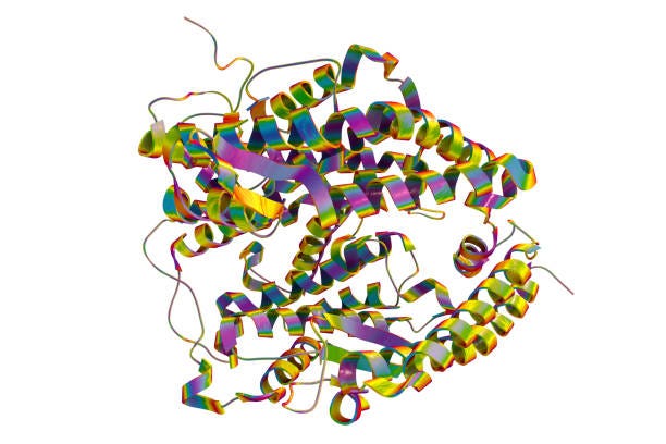
This of course is NOT my focus or interest at all, and when I say that especially “Bio”Chemicals are a hoax and “Do not Exist” this is far from the point I am trying to make. With “Viruses” we have beyond reasonable doubt proved they do not exist as the physical particles they called “Viruses” were indeed mislabeled commonly occurring cellular debris. With “Bio”Chemicals such as proteins there is even less there tangibly to start with, with their very claim. There is no “Protein” particle, you can’t just zoom in a see it with any microscope, the only thing that exists in their claim is a tangled incoherent Computer generated cartoon, as seen below, which is claimed to be the infamous Ace 2 Receptor.
So what do I exactly mean when I say something inflammatory like “Proteins don’t exist”? To answer this question you must be au fait with laboratory processes and exactly WHAT they are claiming is a protein. This is roughly the same with every lab process when it comes to Biology so applies to all. The basic premise that the fraud of Chemistry has set out to do is to refine everything in the Biological realm down to its core components, and laudable task, if correct. The problem is that very soon into their endeavor a bunch of shit stain investors like the Rothschilds and Rockefellers usurped these initial observations of ironically Cutting, Burning and Poisoning everything in a test tube. They pushed it into the realms of the unseen, in molecular and atomistic chemistry.
They ended up convincing the general population that the core essence of a Lemon was Vitamin C that this chemical was the building blocks of what made a lemon, lemony. Now this is a dumb and completely useless trick, take anything, a football for instance, pop it to remove the air, burn it into a puddle, pour some acid on it and finally separate off the liquid with ethanol. What you have left is “The Essence Of Football”, If you drink it everyday you will play more like Messi or Ronaldo (Delete where applicable). Of course this sounds absolutely fucking insane, why wouldn’t it? But this is EXACTLY what they are claiming with vitamins and “Bio”Chemicals. In fact it is 10 times worse, because in today’s barbarically stupid Scientifically Brainwashed cult society we live in, they don’t even get “Essence of Lemon” from Lemons any more, it comes from High Fructose Corn Syrup SMH:
So that is what I mean when I say “Proteins” don’t exist. Of course they have a liquid in a test tube that they have put a label on it saying “In Dis Toob iz ProTeenz”. That liquid has its own properties of usually being a colorless odorless liquid with a distinct pH (Very Important) and contains residuals of the harmful chemicals used to “extract”. It is a Frankenstein soup that causes a set of Lab Values that they claim represent the existence of “Protein”. As I will demonstrate in this article the reagents that go into the Lab assays are certainly not specific to “Proteins” and indeed the environment based on pH, temperature and concentration are the determining factors of those lab values. So the only thing that enables the Chemists to say “This here is Protein”, are the circular reasoned lab values and is entirely just a question of altering the reagents to get the presupposed value they wanted.
Hence with all of the above, there is nothing there, “Proteins” don’t exist in the physical realm as a particle with distinct properties of their own, they, like all other “Bio”Chemicals are just an assortment of random chemicals and processes labelled according to their lab value. I could do the same with anything, labelling a test tube full of Gravy, spit from a camel, toe nail clippings from a Marmoset and Lilt as an “Ace 2 Receptor Protein”, it doesn’t make it so.
So, without further ado and anymore pandering to the intentionally obtuse…. let’s pull this thing apart:
The Beginnings of “Proteins”
1) Who “discovered” proteins?
Antoine Fourcroy (late 1700s)
He and others noticed that animal substances like egg white, blood serum, and muscle behaved similarly in experiments.
But they didn’t have the concept “protein” yet.
Gerardus Johannes Mulder (1830s)
Mulder analyzed substances like albumin (egg white), fibrin (blood clot material), and casein (milk).
He concluded they were built from a similar “core” chemical composition.
Jöns Jakob Berzelius (1838)
This is the key naming moment:
Berzelius suggested the word “protein” (from Greek proteios, meaning “primary” or “of first importance”).
So he didn’t discover them experimentally, but he basically coined the concept as a fundamental biological substance.
✅ If you want one “official” credit:
Mulder characterized them chemically, Berzelius named them.2) How were proteins first isolated?
Early protein isolation was basically kitchen chemistry + precipitation.
A) Egg white (albumin)
People noticed egg white:
coagulates when heated
forms a solid when treated with acids
That coagulated solid is mostly denatured albumin — an early crude “isolation.”
B) Milk (casein)
If you add acid (like vinegar) to milk:
it curdles
the curds are mostly casein
That’s also a crude but real protein isolation.
C) Blood (fibrin)
Blood left to clot produces:
a fibrous solid network = fibrin
this was one of the earliest proteins studied because it’s so visible.
So looking into the Naive interests of our lab scientists a couple of hundred years ago, they pointed out that milk curdles if you put acid in it and if you heat up an egg the white goes solid. I mean hardly rocket science and why you should conclude anything about this property any more than you can cook an egg or make cheese from milk is not clear (Well it is, it is all a money making exercise in propaganda for the state). But here we are, they used Acid which literally changes the charge of claimed molecules causing them to clump, this is the same with lots of different types of “Bio”Chemicals including Sugar which clump and precipitate in acid. This is also the case with claimed “DNA” which later on you will find there is nothing separating these two things really.
How to Test For “Protein”
They are going to tell you that there are chemical assays and dyes that test specifically for protein. These same dyes such as Coomassie Brilliant Blue G-250 are used right the way across the board from simple presence tests that you might have done in school with the Biuret test which is roughly the same stuff, right the way up to Western Blotting in Gel electrophoresis considered the most accurate (apart from claimed molecular tests).
So let’s have a look at this claimed universal “test”:
1️⃣ Who developed it
Developed by: Marion M. Bradford
Year: 1976
Purpose: To create a fast, sensitive, and simple assay for measuring protein concentration in biological samples.
Before Bradford’s work, assays like the Lowry method were accurate but slow and cumbersome, requiring multiple reagents and long incubation times. Bradford wanted something rapid, reliable, and less reagent-intensive.
2️⃣ How it was developed
Bradford was studying protein-dye interactions in his lab.
He tested several triphenylmethane dyes for their ability to bind proteins.
He found Coomassie Brilliant Blue G-250 worked best:
Bound rapidly to proteins
Produced a strong visible color change
Could be measured spectrophotometrically at 595 nm
He optimized conditions for:
Acidic environment (low pH ~1) → stabilizes dye-protein binding
Rapid color development (within ~5 minutes)
Linear range for protein concentration (~1–20 µg/mL)
✅ The Bradford assay became extremely popular because it was faster and easier than Lowry or Biuret methods.
1️⃣ Who invented Coomassie Brilliant Blue
Coomassie Brilliant Blue is actually not invented by a single biochemist — it was synthesized as a textile dye in the late 19th/early 20th century.
There are a few variants: G-250 (used in Bradford), R-250, R-350.
G-250 was later adopted for biochemical use because:
It is stable in acidic solution
It has strong visible color changes
It interacts with proteins via electrostatic and hydrophobic interactions
✅ So Marion Bradford didn’t invent the dye; she repurposed an existing textile dye for protein quantification.
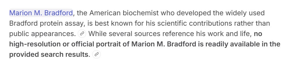
Interestingly enough, both Grok and Chat GPT thought that Marion Bradford who developed this Universal test for protein was a woman, yet on Google search it seems to suggest it was a man?! Even more intriguing is that there seem to be no photos of this person in existence which considering they were claimed to only have died in 2021 at the age of 74 is exceptionally fucking weird.
The other thing that stands out like a sore thumb with this weird story is that this androgenous anonymity supposedly just happened across a Textile Dye used to make blue knitting wools, somehow this bright spark thought hmmm…. I bet that Wool dye will be able to adequately tell whether there is a protein in something or not. Took some into his/her lab and hey presto it worked, what were the odds! I mean call me a cynical bastard but this isn’t really adding up here.
But nevertheless the way this came about is irrelevant, the claim is here that this wool dye test SPECIFICALLY for “protein”.
It’s all just charge (surprising huh!)
As seen above, the Bradford Wool Dye MUST repeat MUST be done in an acidic environment. Without this pre-requisite there is no test. This is quite a major flaw in a test as by definition this then becomes an independent variable in and of itself. Are you really measuring the sample? Or are you merely measuring the pH ( what do you think I think? lol).
So let’s chuck some basic questions at Chat GPT and see if this test passes muster, or will it crumble at the slightest of investigation?
What happens if you add a neutral or basic sample
If you add a solution with high pH or strong buffer, it can partially neutralize the acid.
This shifts the dye toward the green or blue forms even without protein, causing apparent color change.
⚠️ This is essentially a pH artifact.
The Bradford assay is optimized for small sample volumes (so that pH doesn’t shift significantly).
If the sample is too alkaline or high volume, the free dye will turn blue independently of protein → false signal.
3️⃣ Practical lab implications
Samples must not drastically alter the pH of the acidic dye solution.
If your sample is basic (e.g., NaOH, Tris buffer at high concentration), you need to:
Dilute it sufficiently
Or neutralize/dilute the buffer to prevent pH shifts
Otherwise, you risk a false positive.
4️⃣ Key insight
✅ The blue color normally in the assay is caused by protein binding, not pH change.
⚠️ But if the sample significantly raises the pH, you can artificially induce the blue form without any protein, which is why pH control is critical.
In other words: the assay measures protein, but it assumes the acidic dye environment is maintained. Any strong pH-altering sample can interfere.
Well, what did you expect? Of course at the slightest of scrutiny this test crumbled by the wayside. If the sample you are adding is in anyway Alkaline then this test will test positive “without any protein added” in the words of Chat GPT. There we have it Ladies and Gentlemen another notch in the belt, a theoretical Control Experiment win, the test for Protein is actually a test for pH. But it gets better!
Q: The blue color is actually technically from increased anions correct? Short Answer
Yes — the blue color comes from the dye shifting to its anionic form, which is stabilized when it binds to protein.
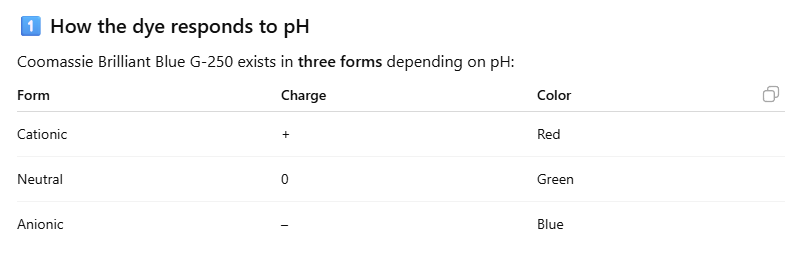
As discussed a few times on this channel, pH is just a measurement of charge. They readily admit this in all literature that the test is a Potentiometer (Measures Voltage potential) of the amount of Hydrogen Ions in a sample. High Positive charge = High Hydrogen Atoms = Acidic and vice versa.
So this should be an easy one to join together, and clarified with AI, that YES the test for a Protein used in every major lab worldwide IS a test for Ionic content or Charge! I obviously proved the same thing with the PCR and implied that it would be the same given that all of these “Bio”Chemicals are measured in Gel Electrophoresis (Which we will discuss in a bit more depth in a bit). But still, it is great to have further confirmation that the exact “Specific” test for “Proteins” is no different.
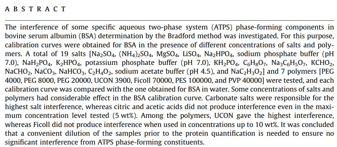
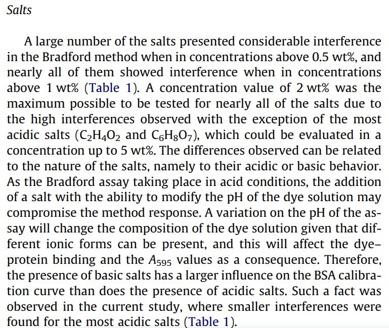
Below is a Published Paper, one of many on the subject of Salts interfering with the Bradford Wool Dye assay. With molecular chemistry they claim that a salt will dissociate into its respective ions in solution, so one would imagine that this is what is causing the interference. One would have to bear in mind that if you are to take at face value Molecular Chemistry that a salt is Ionically balanced so in theory the solution stays the same charge, with each set of Ions balancing each other out. I experienced this whilst conducting PCR control experiments, because I had researched it measuring charge I was searching for substances that would impart charge into a sample, so put all manner of salts in.
This however tended to do the opposite of what I was hoping that samples of things that were giving positive signals were being damped by the addition of salt. So really these types of papers are not even fully controlling for the hypothesis they are really meant to be testing. This is a difficult thing to produce by the way, some sort of “charged” substance because you have to make a very complex system of charged filters so that ions don’t balance out into a salt.
And herein lies the rub, that it is very difficult to “make” a biological sample, one that is “out of balance” that is charged in a way due to the body bringing itself into balance by detoxing the excesses that are “measured” in PCR.
Nonetheless these papers agree that the Ionic components do cause a considerable effect on the Bradford Wool Dye test. so there you have it…
Labelling the “Spike”
One of the few criticisms (that were possible) raised against the Microscopy work where we falsified the morphology of 27 different “pandemic and epidemic causing viruses” was that there was no Immunological staining done on the samples. The idea already presupposes that, as we have found out there are particles that share identical morphology and visual appearance to 27 types of “virus” so may only be separated by extraneous chemical assays or staining. This admission really is confirmation that “viruses” do not exist, as anyone using this line of reasoning has actively thrown out the only direct evidence available in favor of indirect inferences. It is the equivalent of admitting that the CCTV evidence of a murder occurring isn’t evidence and instead choosing to imprison somebody based on the Imprint of a size 9 Nike Trainer footprint.
These inferences when “genetic” are completely extraneous to the sample in Microscopy, but at least Immunolabelling takes place in the visual realm. It is entirely impractical and very expensive to have done 27 different samples with 27 different stains and also poses the issue of not being able to blind an independent CRO to the aims of the experiment, which is pretty crucial in achieving unbiased work and maybe even getting the work done in the first place.
Nevertheless if the opportunity had arisen to do this on the control samples, just for the “spiky bits” of “Sars Cov 2” of course I would have wanted to have a look. This is largely because I have seen what a cacophony of shit the published/peer reviewed papers look like:

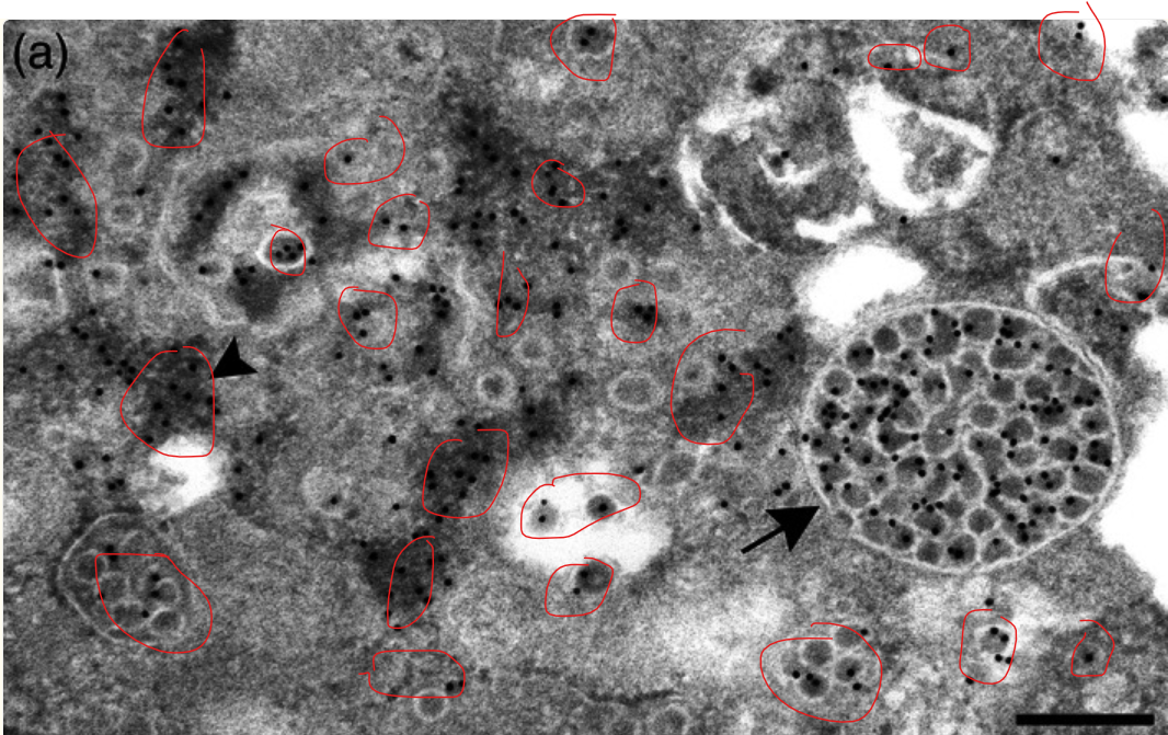

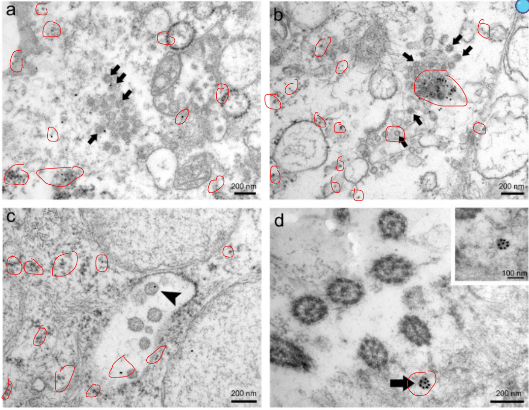

Above represents a couple of papers, most of the images coming from one entitled “Just seeing is not enough for believing” where once again they hold their hands up to morphologically identical particles existing in uninfected cultures and their reliance on these extraneous inferences. The problem is, as with even a cursory glance at the images, this supposedly “Specific” stain that is going to bind only to the “Spiky Proteins” of “Sars Cov 2” to differentiate them from cellular debris is well… the opposite of “Specific” it is indeed completely fucking random.
As highlighted in the red circles I have drawn the Gold Sputtered stain that was supposed “glued” to the back of an “Antibody” that is claimed to ONLY attach itself chemically to “Sars Cov 2 “ proteins. These black dots appear all over the sample near things that can’t possibly be a “Sars Cov 2” virion, floating in dead space in vacuoles and littered amongst cellular debris and quite astoundingly NOT on some of the particles with Arrows pointed at them claiming they are the “Sars Cov 2” particles.
Q: in those pictures the gold particles appear in lots of places not just specific to claimed sars cov 2
Exactly — and that’s an important subtlety in immunogold TEM: the gold particles don’t always bind perfectly specifically.
Here’s why you see them “everywhere” sometimes:
1. Background / non-specific binding
Even well-blocked grids can have some gold particles sticking randomly.
Proteins, membranes, or grid surface sites can attract gold-labeled antibodies non-specifically.
2. Antibody cross-reactivity
The primary antibody might bind other similar proteins, not just spike.
Or the secondary antibody can occasionally stick to other Fc regions.
3. Clustered vs diffuse labeling
Gold is easiest to interpret when you see clusters localized to known viral structures (like the surface of virions).
Single scattered particles are harder to assign as “true spike labeling.”
4. Signal-to-noise issue
Immunogold TEM inherently has some noise because it relies on individual antibody-gold events.
That’s why publications often show multiple virions or statistical analyses to prove the gold labeling is genuinely spike-specific.
✅ Key takeaway:
Immunogold TEM can highlight spike protein, but seeing some gold in “unexpected” areas is normal. That’s why researchers usually quantify localization and don’t rely on a single particle as proof.
Above, I pointed this out to Chat GPT by attaching those images and saying that the labels were completely random. To which it agreed and casually said “Oh there is non specific binding too” !!! SHM. I mean here is absolute proof that the virologists and Mainstream were just lying when they said we needed to differentiate the cellular debris from claimed “Viruses” because only this type of staining would reveal the truth. They can’t even demonstrate this in their own studies. To add to this, of course there aren’t any uninfected controls of the same cell line to prove any of their claims, but really they are not needed, anyone with half a brain can see this type of staining verification is completely fucking useless and it astounds me that the mainstream virologists can’t see that it is actually detrimental to their case being so unspecific in their own papers. But then, stupidity knows no bounds.
Antibodies and the dangerous SPIKE!!!
To make the “Antibodies” (A whole other article in the pipeline) to “glue” the gold onto for the very NON specific staining, they of course need to perform a nice bit of animal torture. They inject what they believe to be “Proteins” into a defenseless animal, wait for a bit until they consider this injection has done its magic and the animal has mounted an “Immune Response”, then they do a large blood draw on the animal and declare they have “Antibodies”.
Now I want to caveat this with a huge warning, I am in no way advocating that injecting a random chemical soup made of strong acids, solvents and blood is NOT harmful, indeed injecting ANYTHING is bad for you and should never be done. However, the entire premise of making Antibodies comes with the idea that they keep the animals well and alive and certainly strong enough to mount what they consider to be an “Immune Response” which is in their minds a mark that the animal is appropriately “defending” itself, something that cannot be done in very sick animals.
When reviewing the literature on these things, certainly when it comes to the smaller animals, these sick bastards obviously kill the animals after they are done torturing them, but in many many studies they keep these animals alive and some even release them. They ALL, in every case note that there are no “harms” bought to the animals. This should clearly come with a large pinch of salt that we know how these sickos operate and it is well within their remit to just lie. But in general the entire premise IS to keep the animals alive.
Therefore when you have the usual Con-Op household names selling their quack “Spike Detoxes” and the numpties running around about “Vax Shedding” they are clearly and provably by their own mainstream science they hide behind are talking verifiable bollocks. There is certainly no biological entity to speak of, it literally does not exist. For sure the chemicals in the Vax are gonna fuck you up, the solution is just not to inject a bunch of unknown shit into your body for a non existent thing. Cheers!
🧬 1. Are animals injected with spike protein?
Yes — in traditional antibody production, an antigen (like recombinant spike protein or a portion of it) is injected into an animal to trigger an immune response. The animal’s immune system then produces antibodies that recognize that protein.
The antigen can be full‑length spike, a subunit (e.g., S1 or RBD), or a synthesized peptide.
Sometimes the antigen is conjugated to a larger carrier protein to make it more immunogenic.
🐁 2. What happens to the animals? Do they get sick or die?
The process is designed to not infect the animal with SARS‑CoV‑2. They are injected with pure protein antigen, not live virus. This protein alone does not cause infection.
Whether the animal becomes ill depends on how well tolerated the antigen and adjuvant are. Usually, animals don’t get sick from the antigen itself.
After repeated injections over weeks, blood is drawn to collect serum. Some labs may euthanize the animal at the end to collect its spleen for monoclonal antibody production. This is a regulatory and ethical issue — in many countries it must be done humanely and justified scientifically
Q: I was looking for a paper that injected the spike protein for the purpose of making antibodies
Yes — here’s a real peer‑reviewed paper that fits what you’re looking for: animals were immunised with SARS‑CoV‑2 spike protein (or spike domains) to produce antibodies, and the study clearly reports antibody production without harmful effects noted in the animals.
🐰 Example: Spike protein immunisation in rabbits
Paper: Antibody signature induced by SARS‑CoV‑2 spike protein immunogens in rabbits
In this study, researchers injected rabbits with purified SARS‑CoV‑2 spike protein antigens (various domains like full‑length S1+S2, RBD, etc.) to evaluate antibody responses.
The goal was to characterise the antibody response, not to infect the animals with live virus.
Rabbits developed strong antibody titres against the spike protein after immunisation, which were analysed for binding and neutralisation properties.
Importantly, the paper does not report adverse health outcomes or harm in the rabbits; immunisation protocols in such studies typically include monitoring for clinical signs, and lack of reported harm implies the animals remained healthy throughout the protocol.
Title: Antibody signature induced by SARS‑CoV‑2 spike protein immunogens in rabbits (Ravichandran et al., Sci Transl Med)
— Researchers immunised rabbits with spike protein antigens to study antibody responses.🐭 Another example: Spike protein immunisation in mice
Paper: Immunogenicity of SARS‑CoV‑2 trimeric spike protein vaccine in mice
Mice were immunised multiple times with recombinant SARS‑CoV‑2 spike protein plus adjuvants.
The study measured high levels of serum IgG and neutralising antibodies, demonstrating a strong humoral immune response.
There are no reports of serious adverse effects in the mice beyond normal, expected reactions to vaccination.
🐇 Context — What these studies generally report
Immunisation studies of this type typically note things like:
body weight, behaviour, and injection‑site checks
immune responses (antibodies, neutralisation)
no significant clinical illness or loss in animals
When no harmful effects are observed, authors do not report disease or mortality, which in scientific animal studies is taken as explicit evidence that the immunisation was well‑tolerated under the conditions tested.
PROTEOMICS IS ACID
In one of these GOF’ed “Virus” papers that was analyzed in a previous article there is yet another massive howler, relating to proteins. In this paper it explicitly says that the indications of this supposed Protein were non existent when put in the completely pH neutral environment of Phosphate Buffered Saline. However as soon as the environment was Acidified all of a sudden these supposed signs of proteins started to show.

Just to fully clarify what is going on here. Acidity is a measurement of POSITIVELY charged Hydrogen Ions. Without wishing to go down another rabbit hole (Which I will do in the future(when I have 5 fucking spare minutes)) into the realms of elemental chemistry; again this is JUST a measurement of Charge. So what they are doing here is very obvious to see when you realise WHAT they are testing for at its fundamental level. They are measuring for CHARGE with either Electrophoresis Banding OR with Chemically charged and Polar Dyes and Stains.
When they put the sample in CHARGED acidic environments, the measurements of CHARGE all of a sudden register. Hopefully you see how fucking stupid this is and can somewhat demystify another complex area of bafflement used in “Bio”Chemical assays.
To note, there is quite a specific window where the pH is just right to get the desired effects. This further shows that there is a customizable calibration for these assays, just like PCR, where it will only amplify where these certain conditions are met, obviously combined with the computer thresholds on the input.


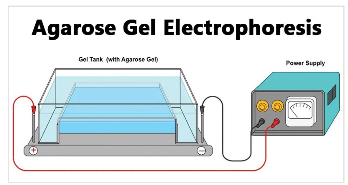
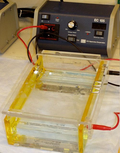
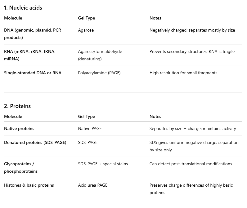
GEL ELECTROPHORESIS
It has been almost flogged to death the amount of times I have mentioned Gel Electrophoresis, but it really is a very good visual aid for those visual learners. What could be an easier way to understand the stupid fraud behind really the entirety of “Bio”Chemistry than seeing the absolute Pony equipment used to measure them. It is a jelly in a plastic dish with a couple of car battery terminals stuck at each end, you put a sample and a dye in and see where the smears stop and declare “SCIENCE HAS OCCURED”…. SHM.
There is literally no better and more visceral way to fully understand that ALL they are measuring is the charge of a sample. In fact it would be INCREDIBLY hard to conclude anything other than that, why it would take a couple of hundred years of brainwashing, an industry worth some Trillions of Dollars and an entire education system operating an aggressive religious style cult to convince people there was more to it than that.
Who was doing electrophoresis before gels?
The big name is:
✅ Arne Tiselius (1930s)
He invented moving-boundary electrophoresis, which let scientists separate proteins in solution (no gel).
This was the first really powerful electrophoresis method used in biochemistry.
Tiselius used it to separate serum proteins and show clear fractions like:
albumin
globulins
He won the Nobel Prize (1948) partly for this work.
Q: Walk me through Arne Tiselius discovery
Absolutely — Arne Tiselius’ work is fascinating because he basically invented the first practical electrophoresis system for proteins, long before gels existed. Let’s go step by step.
1) The context: the 1930s
Biochemists could separate small ions easily, but proteins were a mess.
Proteins are large, heterogeneous, and charged, so they move in electric fields, but in solution they diffuse quickly and are hard to detect.
People knew electrophoresis existed as a concept (Reuss, 1807), but no one had used it systematically on macromolecules.
Tiselius wanted a way to quantitatively separate and measure proteins in serum.
So I have put in blue the absolute zinger in the “Invention” of Gel Electrophoresis. They literally admit that the entire premise was based on the fact that they could “Seperate Ions Easily”, OK…. so that is what it is for. They know this and the “Invention” was for the repurposing of this to be used as a giant distraction for a bunch of made up chemicals. And well would you look at that… the giant repurposing distraction got the highest accolade in duty to the State… the Nobel Prize. If you haven’t figured it out yet… yes EVERYONE awarded this title is a fraud that was incredibly useful in painting a made up story to cement a piece of political science for the future generations, and we just saw this hand over in full HD.
As you can see from the table below, showing all of the “Bio”Chemicals that are measured using Gel Electrophoresis, it is practically all of them, below this table is a list of the ONLY “Bio”chemicals not officially measured, usually not because they intrinsically can’t but more because they would be deemed inaccurate because they are claimed “too small”. This list of “can-nots” is really just glucose, vitamins (don’t exist), some hormones (don’t exist) and fats. So Sugars and Fats which demonstrably exist by their physical properties on the macroscale so need not to be “Gelly Zapped”.
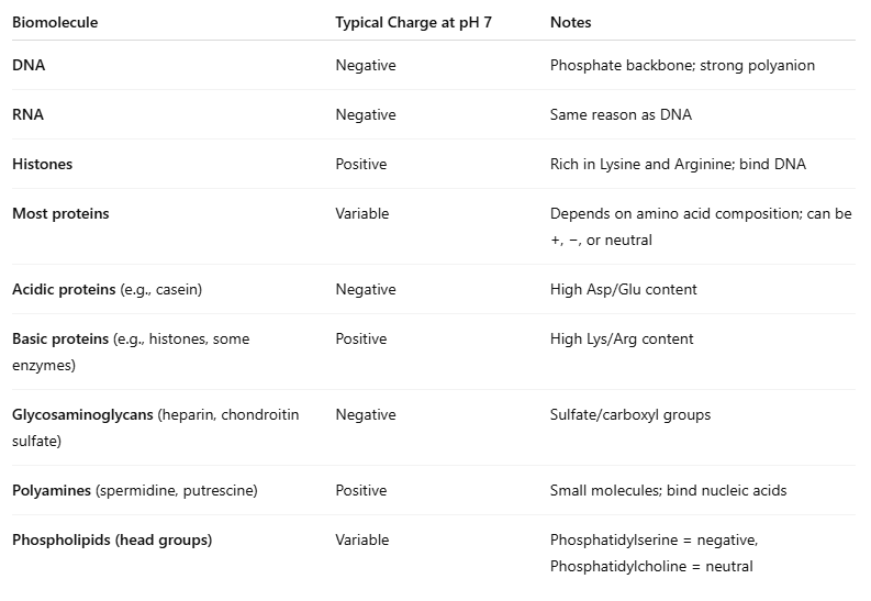
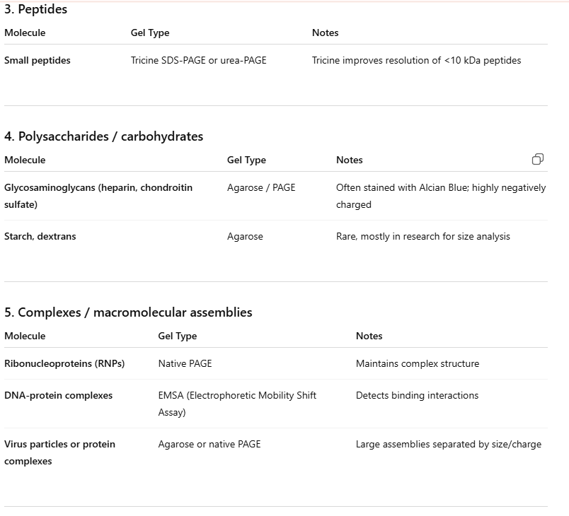
Biochemicals that generally cannot be measured well by gel electrophoresis
1) Small molecules (most metabolites)
Things like:
glucose
ATP
vitamins
They’re usually too small and don’t stain well.
2) Steroid hormones
Like:
testosterone
progesterone
They’re small, mostly nonpolar, and don’t behave like proteins/DNA in gels.
3) Lipids
Like:
triglycerides
cholesterol
phospholipids
fatty acids
They’re hydrophobic and don’t migrate in normal gels properly.
As above with the interference of Ions in Bradford’s Wool Dye assay, there is obvious interference from anything charged such as salt dissociation in Gel Electrophoresis. Coincidentally, Gel Electrophoresis ALSO uses the dodgy Wool Dye in the first stage to stain the “Proteenz” in the first place. So we have compound interest on this thing of it only testing charge to stain, then only testing charge in the gel. I would call it a house of cards, but it is literally only 1 card standing on its end, they are looking at whether it is the back or the front of the card and the rest is just all made up.
Q is coomasie Blue used as a dye in western blotting?
Yes — Coomassie Brilliant Blue is used in western blotting, but not as the main detection dye.
What it’s used for in western blots
To stain the gel (before transfer) to check you actually loaded protein
To stain the membrane (after transfer) as a total protein stain / loading control
CONCLUSION
We had all sorts of weird and wonderful information cropping up that puts a Steak in the heart of the “Proteen” narrative. From the extreme oddities of our Androgynous, Anonymous Marion Bradford, who stumbled across the luckiest discovery known to man when he/she was walking between her/his wool dying shop and laboratory and happened to be carrying an open cup of Coomasie Blue textile dye and WHOOPS! tripped and fell and it landed on something he/she knew that had some but not all protein and miraculously it only died the things he/she knew was protein (before the dye was know about for this purpose).
With such luck and such an important role in history for being the person who made the assay that worldwide would confirm the presence of every protein known to Mankind, they died a mooted death just 4 years ago, having never repeat NEVER having a single photo taken of them, nor any sort of descriptive information confirmed about them to the point where nobody knows whether they were a bloke or a woman.
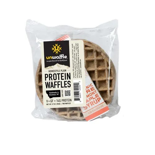
We have seen right across the board from the wool dye to the Electrophoresis that this is just a measure of charge with a smorgasbord of environmental tweakable variables that alter the lab value, the ONLY thing that brings our Sharpie Labelled mystery liquid into existence. No, this is all a ruse, the same with EVERY “Bio”chemical a fraud to try and package “Natural” stuff and sell it with endless avenues of marketing: Protein Waffle anyone? I’m sorry but if you can’t see that no, this Belgian Confectionary does not have 33% added “essence of steak” sprinkled onto, I don’t know what to tell you.
I will approach the “rough” end of the fraud of “Bio”Chemistry at a later date in an article I have lined up called the “Bio”Chemical challenge. This sets out the challenge that if you truly believe in all of these “Bio”Chemicals that are the exact refined nutritious sources of the natural world we live in then you don’t need food, you can just live off this shit for ever. This is the visceral control experiment for determining this as has, been somewhat attempted by a fair few (With disastrous results), so stay tuned for that.
We also saw the “get out clause” proffered by the opposition, a cry for IMMUNOSTAINING! Well… what a laugh that was, the greatest self own of non specificity I have seen, possibly in science, it is no wonder there aren’t too many papers out there doing it and offering it as any sort of evidence. Of course under the slightest of pressure the mainstream narrative encyclopedia of AI turned over its cards and admitted its claimed specificity was a massive fucking lie, so we notched that win into the woodwork.
Finally we zoomed in on the baton-hand-over of the “Bio”Chemical fraud, Arne (Hatsa La Vista) Tiselius was given his decorations, his Maltese Cross for services to Scientific Psy-Opery when he created the largest smoke screen repurposing of an instrument working perfectly well for moving Ions from one place t’other and turned it into a Money-Printing-Belgian-Waffle-sprinkling-Wool-Dye tool for the state to write blank checks on medicine, drugs, confectionaries, vaccines and scamdemics based on a bunch of smears in an electrified jelly.
So no, Da Big Bad Spikey bit ain’t up to much, there is no such thing ,obviously, the Mainstream told you this but you just refused to listen. They want to keep these animals alive so that they can drain the poor sods of literally liquid money, because the global Antibody production market is worth $15-17 BILLION (Yes with a B), if they can keep these animals alive and keep harvesting them for their precious liquid, they certainly ain’t killing them on purpose with the “Proteenz” they are injecting in them. So the Spike Protein Story is obvious full frontal codswallop.
So onto the next one in the “Bio”chemical circus, maybe specifically antibodies or potentially hormones, whatever takes my fancy, but I’d wager heavy money it all being roughly the same hoax. Once you see the pattern, the lazy, fat, greying council worker at the Bureau of Scripting Establishment Science never really deviated too far from the same way of cheating observable reality, so we will continue to lift that veil.
Peace./

This article is available as an Ebook on Shadow Banned Library
They want everything We know to be wrong. Thanks for setting a few things right!
took some digging but found a photo https://www.onlineathens.com/story/news/education/campus/2019/10/27/uga-researcher-honored-with-distinction-award/2434565007/