

FLUORESCENCE

This is the third installment of the breakdown of the individual PCR reagents. Today we are going to be looking at Fluorescence, which I find quite exciting (pun intended). Finally in all of this garbage we have come to the business end of the deal, the tangible, visceral thing that happens and is measured in a RT-qPCR reaction. Fluorescence is certainly a thing, of course we can SEE it, it can be quantified and measured and predicted with high precision, this is not really debatable.
The *Light Display* really is the favorite show-stopping magic trick of the Chemist. One should probably, if they had a cliched experience of education, have been taught Chemistry by an eccentric pyromaniac preferably with long singe-able eyebrows. My Chemistry teacher was no exception to this rule, Dr. Cook, a jumpy, twitchy old man that could never quite, at any one time, get all of his collar to sit down on his lab coat. He would sit there in grey tedium until his number was rung and it was time to get the plexiglass screens out for “Demonstration Time”.
As his name would suggest he always took the liberty of adding a magic sprinkle of chemicals more than the recipe required so that the resultant bang, whizz, fizz, pop or splurge would just avoid the protective screens and ricochet off the suspended ceiling tiles and into the crowd. Yes this big *something is happening* gives the Chemists full artistic license for WHY the Bunsen Burner flame has turned Green. Really it is this disconnect which is the Wizard of Oz trick, green smoke and mirrors hiding an eccentric Hobbit on a power trip.
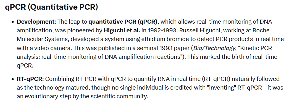
Before we get into the nitty gritty of Fluorescence in general, I want for us all to acknowledge a collective sigh of relief as I give you this good news: We can talk about PCR and not have to mention the name of that gigantic wazzock Kary Mullis- hopefully ever again. The most modern and widely used form of PCR, Real-Time Quantitative PCR actually is quite far removed from the original PCR design, as it uses Fluorescent Probes that are measured with lights and cameras to give in-play readouts. This was invented by a bloke called Russel Higuchi, so from now on in, this guy is going to get all the flack for this fraudulent invention( the prick lol).

So let’s start from the top and look through step-by-step at what Fluorescence is, where it comes from and how it works.
Fluorescence is claimed to be the excitation of certain materials with UV light that releases light at a longer wavelength. A change in wavelength is usually just bought about by a change in medium property/density/opacity like light moving from air to water or passing through plastic/glass colour filters. The “glow” is seemingly just reflection, light bouncing back after being slowed down in a medium change that happens to have a desirable specific wavelength that can be seen. I don’t think there needs to be anything complicated about “electrons and molecular structure, absorbing and releasing energy”.

It seems that quite a lot of just normal materials fluoresce. I did most of my experimental research for this area during my twenties gurning my face off listening to Drum and Bass. You will observe the “natural phenomenon” of white t-shirts and trainers and people’s teeth and eyes and Mistsubishis Fluorescing in the UV strobes. Apparently this is because they use “optical brighteners” in t-shirts with “naturally fluorescing compounds”.. I mean I would need to be quite a bit more mashed than I was then if I were to believe that.
So here I got a list of some of the main Biological Substances known to Fluoresce which includes some pretty amazing finds:

I mean Chlorophyll is in every plant on the planet right…. so that means every plant sample taken for PCR will Fluoresce.
Collagen is an interesting one. Just to point out here it is the most abundant protein in the body. It can be found in saliva and mucus. Mucins also Fluoresce btw. So it seems that practically every human sample will naturally fluoresce. This one is extra interesting for me as I proposed the stringy jizz that is claimed to be DNA from a Strawberry was probably just Collagen in my article here on the DNA Hoax.

Amino Acids like Phenylalanine which is found in nearly every major food group also Fluoresces.
NADH, Ok this is a huge one. Some claimed Enzyme to do with respiratory function. Whilst I don’t think that Enzymes are what they are claimed, they may still be registered in these tests as a “property”. The bombshell here is that this knowingly fluorescent material is elevated and expressed in sick people.

Lignin, so combined with Chlorophyll, basically plants are off the charts in their glowingness.
Retinol (Vitamin A), this will be of great interest to my Twitter friend Dr. Garret Smith who does great work revealing the dangers of Vitamin A and his Toxic Bile Theory (check it out).
As mentioned in the above video that Black-light is often used at crime-scenes as blood, semen and urine all fluoresce. Yes it seems we have a common theme when it comes to piss that it is also a naturally fluorescing waste product.

Bacteria and Parasites
All of the very long list of biological substances and materials that Fluoresce didn’t contain the biggest bombshell of them all, the one that reveals that a lot of Bacteria and Parasites naturally Fluoresce under UV light.
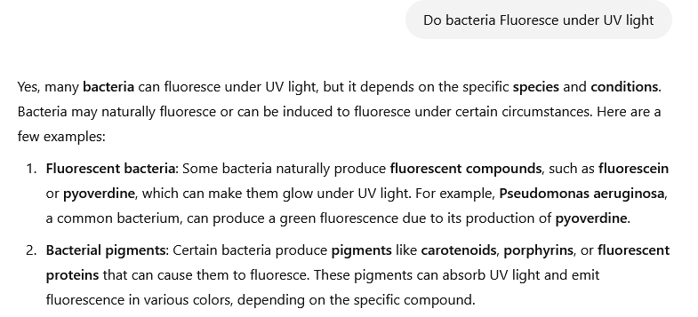

So yes you read that right…we don’t need to deliberate on the existence of Bacteria, they certainly increase in a diseased state as they are the Clean-up crew in our bodies helping ridding of dead and decaying tissue as well as toxins/chemicals/poisons etc. A lot of these Bacteria that Fluoresce under UV light are commonly found in Saliva and Mucus.
This is the same deal with parasites:

So just the natural Oral mouth flora of bacteria, things like Streptococcus Mutans which is the most abundant Bacteria found in the mouth just naturally Fluoresces. OK that is quite a big deal if you are taking fluorescence as a Marker in qPCR, as simple as a sick person has more bacteria in the sample (not causing the disease but a by-product) it fluoresces more.

Watch this video about Bacteria literally exploding in Fluorescent light displays when exposed to UV light. Now this is claimed to be an ever so slightly shorter wavelength (250nm) than is used in qPCR (480nm).
This seems a little unbelievable that the difference between exploding and being perfectly fine in a qPCR reaction is just a tiny fraction if a percentage difference in wavelength. Combine this with the fact that high temperatures, like the very quick temperature changes experienced in qPCR cause Bacteria to “lyse or rupture” and you have yourself a full working theoretical model for how qPCR could be measuring something completely different to what it claims. I would probably caveat this with the fact that I probably don’t believe it is this. Certainly not the MAIN thing that qPCR is measuring, but it could certainly affect the results.

Mechanics of Fluorescence
Some of the initial discoveries of Fluorescence were very simple observations, George Stokes shone a UV light at Fluorite, a mineral, and in glowed and he called it Fluorescence and they gave him a knighthood so his name was now Sir George Stokes. Good science.. no seriously I am happy with that, if science operated as simplistically as this I think we’d all be better off.

Let’s go to the next phase of Fluorescence when they try to extract it.. this time it was by milking a Jellyfish in 1962 by a guy named Osamu Shimomura. Here he is, holding his Jellyfish.


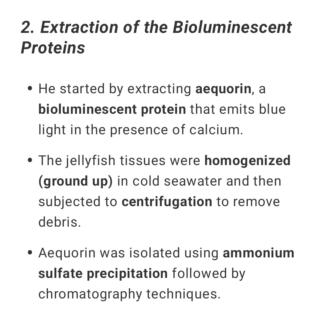
So much alike every other supposed isolation of anything in Biology, what they do it they take an animal with desirable observable attributes, grind it up and then precipitate it out of solution and say “hey presto” we have Pure desirable attribute, in this case fluorescent protein.
The interesting thing about this discovery (for which he won a Nobel Prize(do they just hand these out in cereal packets)) is that they found that this Fluorescent material ONLY glowed in the presence of Calcium.
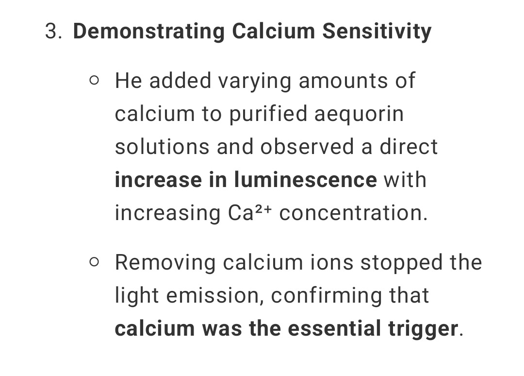
Interesting that it needs a charge ion for the Fluorescence to be seen. Also interesting in my previous article when looking at IVF that eggs will NOT develop, even after sperm is introduced to them unless they have a charge applied, which in nature is done by Calcium ions.
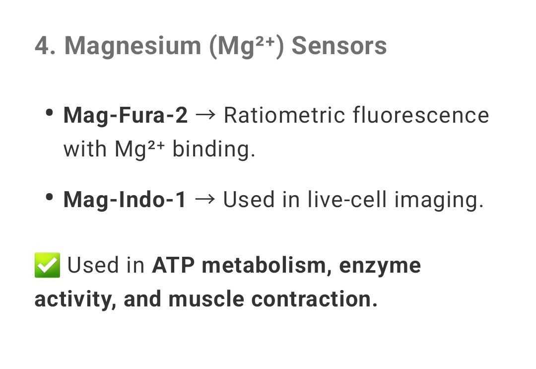
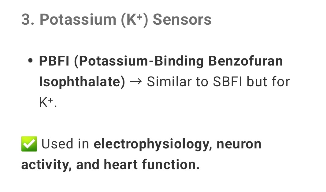
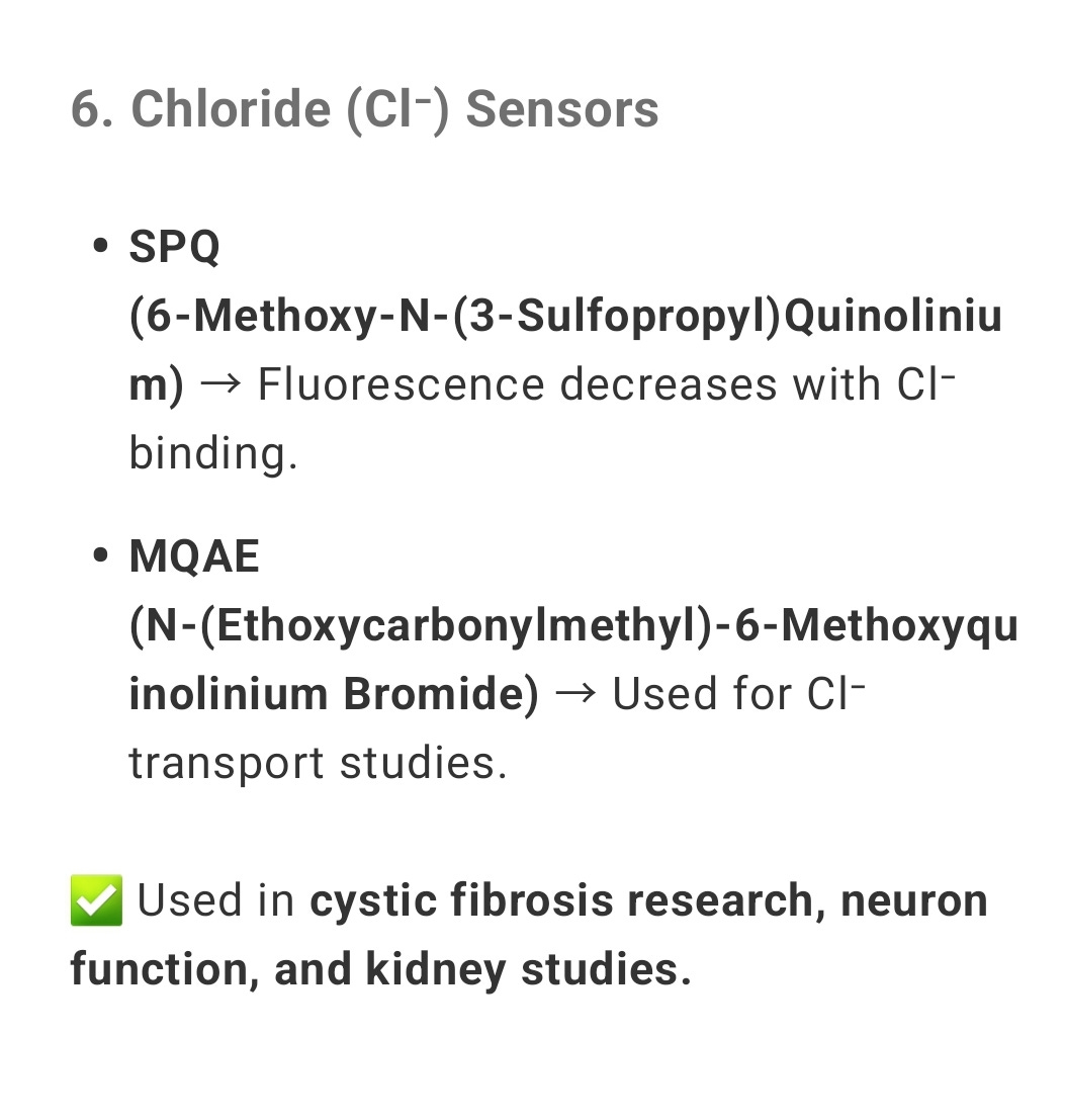
So this effect of only glowing in the presence of Calcium is used to detect Calcium in the body, there are many other different Fluorescent materials that are only triggered by certain Ions so again are used to detect these specific Ions in the body, let’s take a look at a couple of them:
Magnesium
Potassium
Chloride
Also used for pH/Acidity Detection
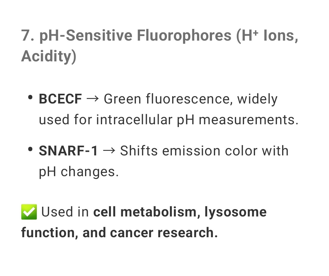
Let’s take a look at an experiment of increasing concentrations of Sulphuric Acid Increasing Fluorescence in Quinine Sulphate. This is literally described in the experiment as the Acidity is giving charge in the form of Hydrogen Ions which makes the substance Fluoresce.
Ethidium Bromide
Before the invention of qPCR and specifically tagging with Fluoresce whilst the reaction was occurring, the old form of PCR done by HeWhoShallNotBeNamed was fluorescently tagged AFTER with Ethidium Bromide, this was to supposedly visualize the amount of DNA produced after replication.
Again the interesting thing here is that it did this based on charge.. Ethidium Bromide is positively charged and so just assumed that is bound with DNA in a sample (Although as we have seen countless times ALL of these things like proteins, amino acids, enzymes, hormones etc etc are all or can be Negatively charged.

qPCR
How does this all fit into PCR I hear you scream… are you ready to enter into the realms of complete fantasy once again, Jeeves, Fire up the animations.
Here we see what they claim occurs in the qPCR reaction, this one is actually for LAMP ; ) .. which uses identical reagents, just with one continuous heating length rather than cycles.
So the principle is that they can attach a Fluorophore to one end of a nucleotide sequence and a Quencher at the other called a Probe. The Quencher is basically a damper for the Fluorescence. It is claimed that ONLY when the correct primer sequence is aligned to its target sequence does an all knowing and totally obedient enzyme come along and “snip” this Probe at exactly the right nucleotide (don’t ask how it knows). This causes the Quencher and Fluorophore to move apart from one another and given that the Quencher only works to dampen the Fluorescence at close proximity, the Fluorophore can now be seen and you get a positive indication on you PCR.
Wow so Tech.

I will end up sounding like a stuck record on this, but I don’t really care, it is one of the complete absurdities of this whole fake reaction and until people truly understand this point ( or you are always welcome to challenge my thinking on this) I will keep making it: All of this is chemicals in solution, to suggest that you can just attach something to one “end” of a liquid, and a something to the other “end” is nothing short of mental. It has no physical shape or form it is just a liquid.
This is even more poignant when you look at the entire premise for what is happening with the Quencher. It is claimed that the Fluorophore will always be glowing when the UV light shines on it, it is just the Quencher that stops the glow from being seen. They claim that the ONLY reason why this works is PROXIMITY or DISTANCE from the Fluorophore when “snipped”. BUT THERE CAN BE NO PROXIMITY IN A SOLUTION. Even AI agrees with me on this (It then goes off on a completely Invented story about theorized and never seen molecular structure. But Nonetheless, it admitted there is no physical proximity.

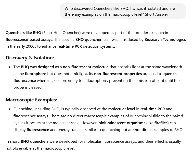
The Quencher used in a lot of these is called a Black Hole Quencher. It was “discovered” in the early 2000 by Biosearch Technologies. The huge problem here is quite HOW they came about this marvelous feat. There are NO Macroscopic examples of this being even possible where a molecule of something just sucks all of the light out of something (like a Black Hole **Bullshit Alert**).
So they developed something on a microscopic level that they couldn’t see, of which there is no know instance of this thing working in real life. It is like saying I am putting Microsopic Flying Pigs in this test tube…
Q: “How do you know they are flying, because they don’t do that in real life”……
A: “Trust me Bro”.
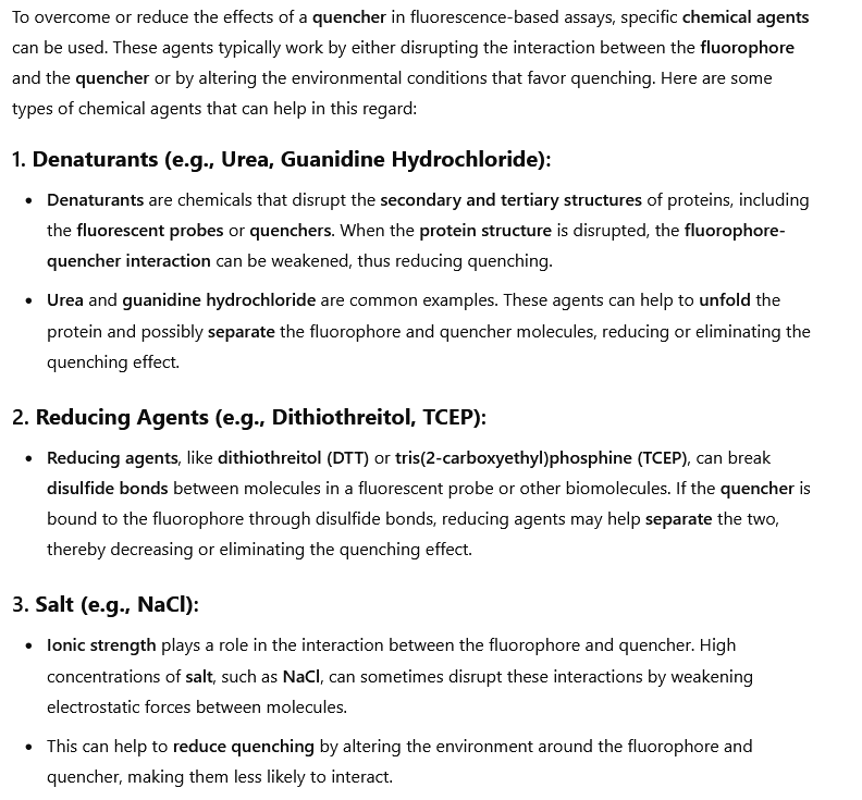
Let’s just assume for the sake of brevity that they have created tiny little Star Trek Quasars and put them into the test tube, what are the limiting factors.. what are the things that reduce the effects of this Quencher that are not “proximity” from the Fluorphore?
Yep…Piss.
You seemingly read that right… I am not making this up or specifically searching for this at all… this research is going to give me a bad name.

An incredibly interesting set of variables, most of which are knowingly put into the the reagents of the qPCR test BTW. They knowingly put salts and Metal Ions in the form of Potassium/Sodium/Magnesium Chloride. We have seen that both Alcohols and some Acids in Soft Drinks make positives on the Antigen Tests.
If we now compare this to all of the factors that affect the brightness of the fluorophore:

We have here a synergy with the limiting or boosting factors of the Quencher, most notably the Metal Ions and pH (ONCE AGAIN). It is becoming an incredibly common theme when I look into the theory of the PCR, at the root of ALL of it is CHARGED particles (as essentially this is what acidity is claimed to be is charged Hydrogen molecules). We also have a few interesting ones, temperature and viscosity. Quite clearly this is a bit of a problem in a THERMOcycler if temperature naturally increases Fluorescence (this is accounted for I presume LOLOLOL). Also Viscosity of the sample seems like a big one… the more sick, the more dehydrated the more viscous.
CONCLUSION
Fluorescence is THE thing that is being measured in the qPCR reaction. We have seen however that practically every fucking living thing in a sample of Saliva or Mucus or Blood Fluoresces from Collagen to Amino Acids to NADH to Vitamin A to Bacteria. What is NOT fluorescing in UV light might have been a better qPCR test.
We saw that a major trigger for Fluorescence is Metal Ions such as Calcium, Magnesium and Potassium. Coincidentally these are also huge limiting factors to the Quenchers (Never seen in real life) that are supposedly blocking out the Glow until something VERY specific happens.
Altogether I hope you can see just how implausible the story is for WHY a Fluorophore lights up in the official narrative, tenuous, complicated and ultimately physically impossible stuff happening as a cover story for basic heating up of Metal Ions in slightly acidic environments.

I am devouring everything I can find regarding voltage gradients and it is clear that they are using FLUORESCENT DYE that is voltage sensitive, as Sally Adee described it (We are electric, p.202) - I have not seen anyone who has made this connection clearly yet ! I think this is what Dr. Ana found everywhere in the nasal swabs, antigen test / pcr (it distributes over the whole face, head and there is UV sensitivity) - this is what is constantly sending light signals from our voltage gradients, to then be able to live read our ion channel communication systems / radiate back - they are literally doing that !
There are fluorescent nanoparticles used for that purpose and they are being found in the environment! Voltage Gradients become a light signal that can be picked up by sensors, regarding the PCR for example (as a possible route to get coherent data from the readings) ...
Ok so regarding PCR - it is the excitement from the light, allegedly, that brings the electrons forth from the "DNA" - but what if it is truly the ionic charge of the organic material being red out through fluorescent dye- that gives both spacial- and temporal voltage information (like in Levins experiment where you can see the electromes activity through voltage sensitive fluorescent dyes on organic material ) ... both is possible, since the exact chemical formula of the dye is not disclosed ....l
Dr. Ana describing the findings around "fluorescent dyes" https://rumble.com/v53x8gg-glowing-c19-shots-and-fluorescent-nanotechnology-with-pfizer-whistleblower-.html
they are making the voltage gradients visible and easy to pick up, very smart (but demented) !
Fun find : "Voltage-sensitive promoters: DNA sequences controlling luciferase gene expression, activated by voltage-dependent cellular events. Example—depolarization opens calcium channels, calcium binds transcription factors, promoter turns on, luciferase produced. Not luciferase itself responding to voltage, but its production triggered indirectly.
Substrate availability tied to membrane potential: Luciferase needs luciferin, ATP, oxygen. Membrane potential shifts (e.g., depolarization) can alter ATP synthesis via mitochondria or ion-driven transport of luciferin. Voltage affects substrate levels, thus luciferase activity, not enzyme’s structure or function directly." so there you have it, it is not luciferase directly that triggers the voltage sensitivity - it is the genes telling it that first and then it is voltage sensitive. But the A.I. denied it first ;)
Amazing work thank you. You’re on a roll. What really has me shaking my head is that one smart guy like you, with some common sense and simple experimentation can demonstrate these truths, and all the PhD‘s, professors, and universities in the world apparently can’t seem to figure it out.