


Appearances Can Be Deceiving - Viral-like Inclusions in COVID-19 Negative Renal Biopsies by Electron Microscopy
The published, peer reviewed control study.

All of the content that is put out on this Substack is going to be for free. If you feel so inclined to donate or Sign up for a Paid Subscription that is very much appreciated. It will keep me writing, putting out content and continuing the largest Control Studies Project falsifying Virology.
Over the past 4 and a bit years myself and a large group of researchers have suffered in debate over the “V” question. We would often encounter a few distinct groups of people, Vaccine/Mask/whole narrative swallowers, Pharma Pushers and Bioweapons from Bat Anus (honestly Jerm!) theorists.
The Full Monty viral believers don’t stay for very long and can be silenced with one simple question: “Have you had a Monkeypox booster yet? If not, why not?”.
The Wuhan Weapons enthusiasts leave shortly after you point out they put their deadly bioweapon up people’s noses and it did nothing.
But the Pharma Pushers, the people claiming to work in labs and have vested interest in all things needles and poison, they unfortunately stick around. Obviously most have a financial incentive like the Johnson and Johnson employed Debunk Da Funk (Aka Funky Danny). But incentive aside, this is where the real battlefield for us was.
Really, how myself and my fellow researchers cut our teeth was going toe to toe with these people exchanging “papers”. Just like any Literature Review, you have to quote and reference papers to back up any claim you make. It is an odd position to be in as most of these “papers” treated as if authoritative “passports” were, to us, complete and utter junk. We would only cite them to show how and why they were wrong in their methodology, either through blatant negligence or more often than not proving themselves to be wrong in their very own paper.
Unfortunately as a group we had to (literally) review thousands, maybe tens of thousands of papers to find a handful that SUPPORTED our claim. I.E not ones that we just cited only because the flaws in the methodology meant we could ignore them, no these papers were so rare you could count them on one hand.
We had to scour Google Scholar for the rare occasion that not only virologists had muffed up and done a control experiment by comparing infected/uninfected samples, but had also gone onto publish the results. This is one of the huge reasons for running this project, is to provide that evidence so that we didn’t have to rely on these, mostly quite old, and unique papers. It would be in our hands, with our interpretation, rather than relying on reading between the lines of the massive cognitive dissonance of a virologist that has slipped up somewhere.
One of those papers was entitled: Appearances Can Be Deceiving - Viral-like Inclusions in COVID-19 Negative Renal Biopsies by Electron Microscopy.
It was the ONLY paper that tackled the area of Electron Microscopy comparing TEM images found in uninfected samples.


So right from the very off the categorically state in no uncertain terms that they have found particles that are “morphologically indistinguishable” from “Sars-Cov 2” not only in samples taken from negatively tested patients but also from Renal biopsies taken before 2020.


Here we already have a problem in that this paper is cross referencing these particles found in negative patients with “Sars Cov 2” with “distinctive 9-12nm spikes”. Yet there are plenty of references of these “viruses” that clearly have no spiky appendages. There is even a category called “bald” to describe these found in “positive” patient samples.
So clearly they are cross referencing and finding an even more specific morphology. Imagine how more uncertain they would be if they were to cross reference the ones without the spikes too.

Another revelation next is that they insinuate that they have never had any positively tested urine sample, hence meaning that there really was no possibility of there being a “Sars Cov 2” particle in their uninfected samples…. A true control!!

Then in possibly one of the most revealing passages of text in a recent virology paper; they not only say effectively that you should not take the morphology of a particle as evidence of infection, implying that there is no evidence of “Sars Cov 2” existing in Electron Microscopy alone. But they also say that the supposed specific antibody tests they used to stain these particles is actually not specific at all, because they can be absorbed by cell tissue itself, such as in the proximal tubes, in this case.
Although we have done no immuno-histochemical control studies yet, it very much seems that we would achieve some favorable results in falsifying the Antibody testing area of virology, especially given that the HEK293 cells we used in the cultures have similar properties to these “absorbent (lol)” proximal tubule cells.


The above section references the below study which is linked through the photo. This is pretty stunning stuff that I hadn’t previously seen in this paper because I didn’t follow the links provided. Here they say that ALL kidneys they have ever tested show “NON SPECIFIC POSITIVE STAINING”.
What are most of the cells used in the culturing of viruses? You guessed it… kidney cells!

So they conducted a review of 15 different samples and found “viral like inclusions” in ALL 15 samples.


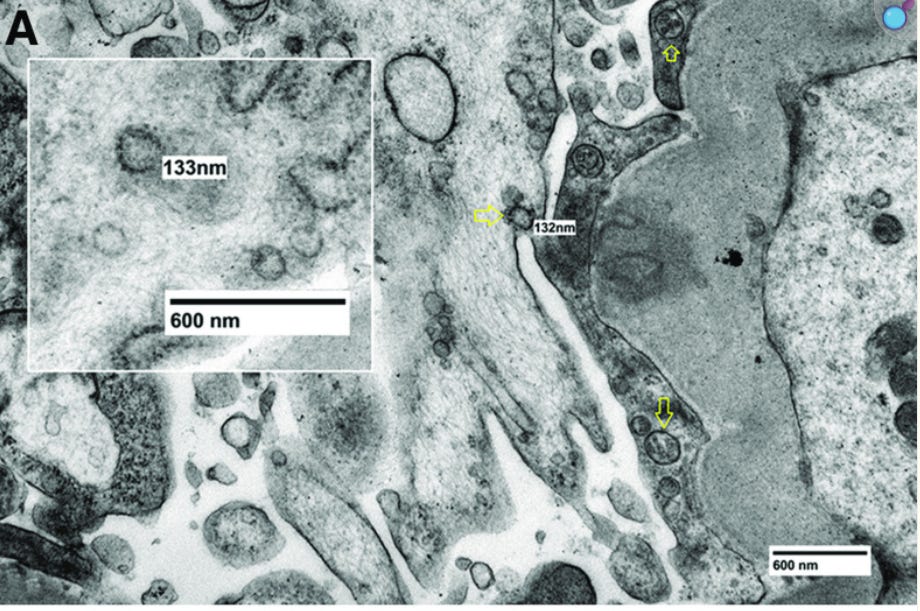

So here are some of the pictures taken from these samples and as you can see… they do indeed look like what they call “Sars Cov 2 Viruses”. They have pointed them out with their yellow arrows, which adds to the effect, they are the right size and in the case of some of the things they call these particles have the identifiable spikes. Although as above there are also versions that they call “Sars Cov 2” which do not have any spikes.
How many “viruses” would they be able to identify if they were cross referencing “viruses” NOT with these distinctive spikes? Well I hope that a lot of the images released from our control cultures starts to answer that question, being I think you could probably find particles that look like every virus ever in the detritus from cell lines breaking down in a petri dish.

They end this bombshell paper with one last zinger. The revelation that they have been finding these “viral-like particles” back from at least the 1970s!! They again note that the only way to actually confirm these things existence is to test them genetically. Convenient that along comes PCR in the mid 80s as the rescue device to stop people from noticing these particles in uninfected cultures do indeed look exactly like the things the virologists were calling “viruses”…

The non scientists either won’t understand the significance of the findings described here or will pretend not to.
The virologists will contemptuously say “You just don’t understand anything” and will insist you’re not following the scientific method. Their yap dogs will repeat it.
Meanwhile, we know exactly what it means.
There’s no scientific evidence for the existence of viruses.
I like that phrase people use when telling a story; “you can’t make this shit up.” But wait, they did make it up. The whole science of virology made up from whole cloth. The best part is their own papers disclosing the truth. Thank you, Jamie, for reading them.